- Refraction
TERA
The Topcon TERA combines advanced Placido-based corneal topography with a comprehensive Dry Eye Suite – designed to support confident evaluation and management of ocular surface disease. Its automation, precision, and flexibility make it ideal for ophthalmologists, optometrists, and specialty clinics alike.

Key Features
 5MP High-resolution Colour Camera
 Fully Automated Topography,
Pupillometry and NIBUT Tear Meniscus Height
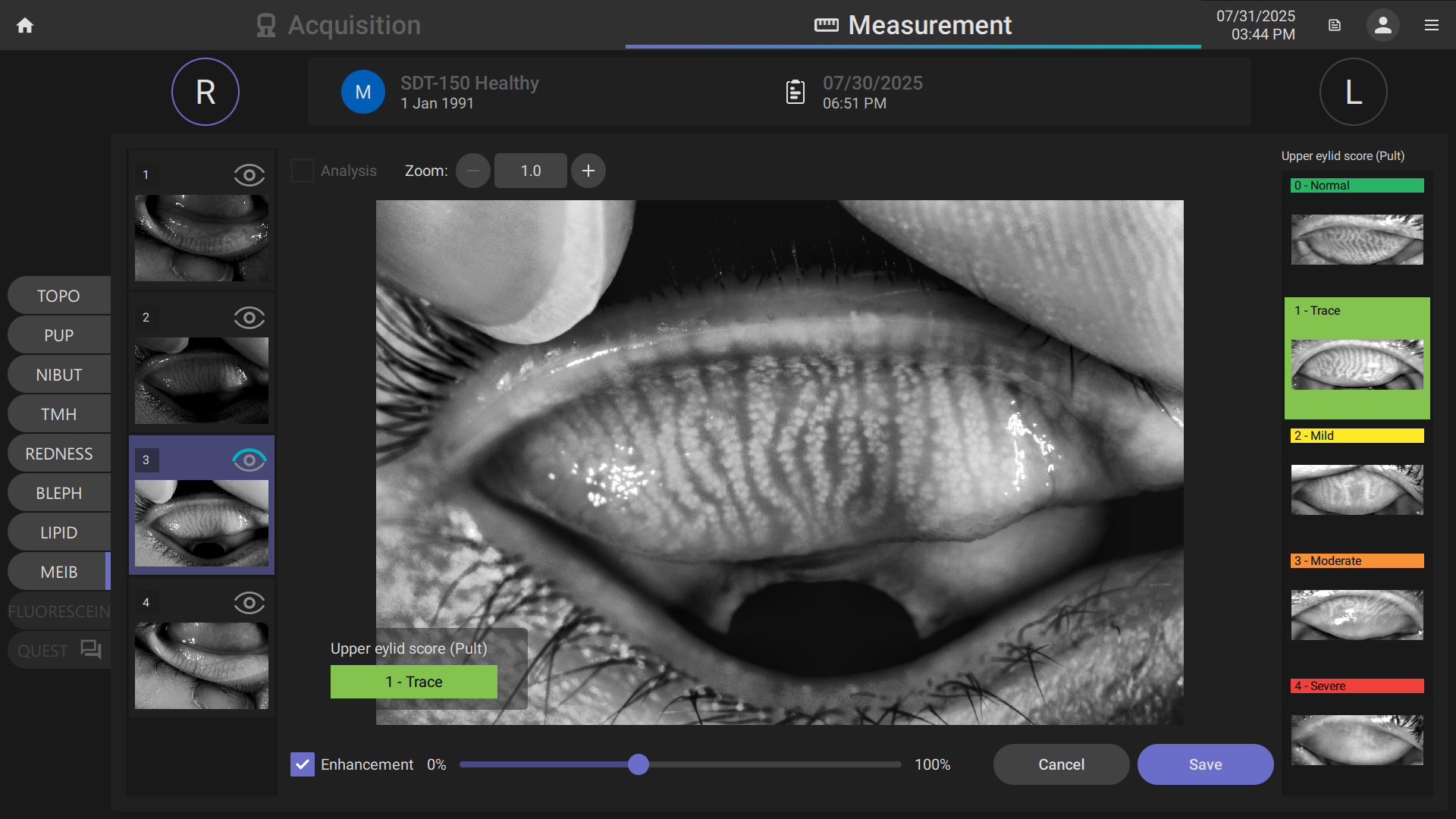
 Meibography
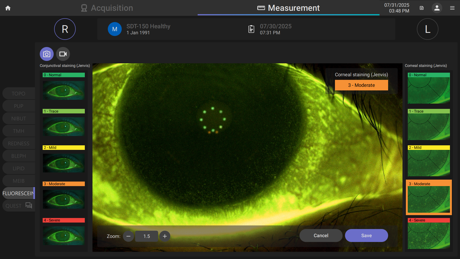
 Fluorescein Staining Assessment
 Blepharitis Assessment
 Lipid Layer Assessment
 Conjunctival Redness Assessment
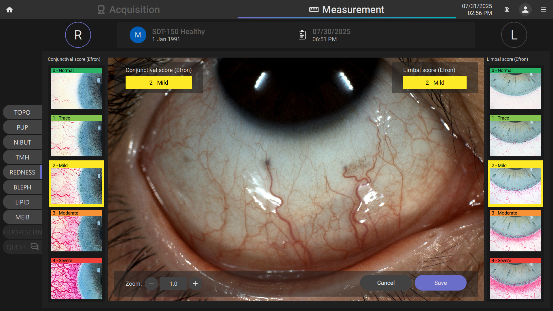
CLEAR VISION FOR DRY EYE MANAGEMENT
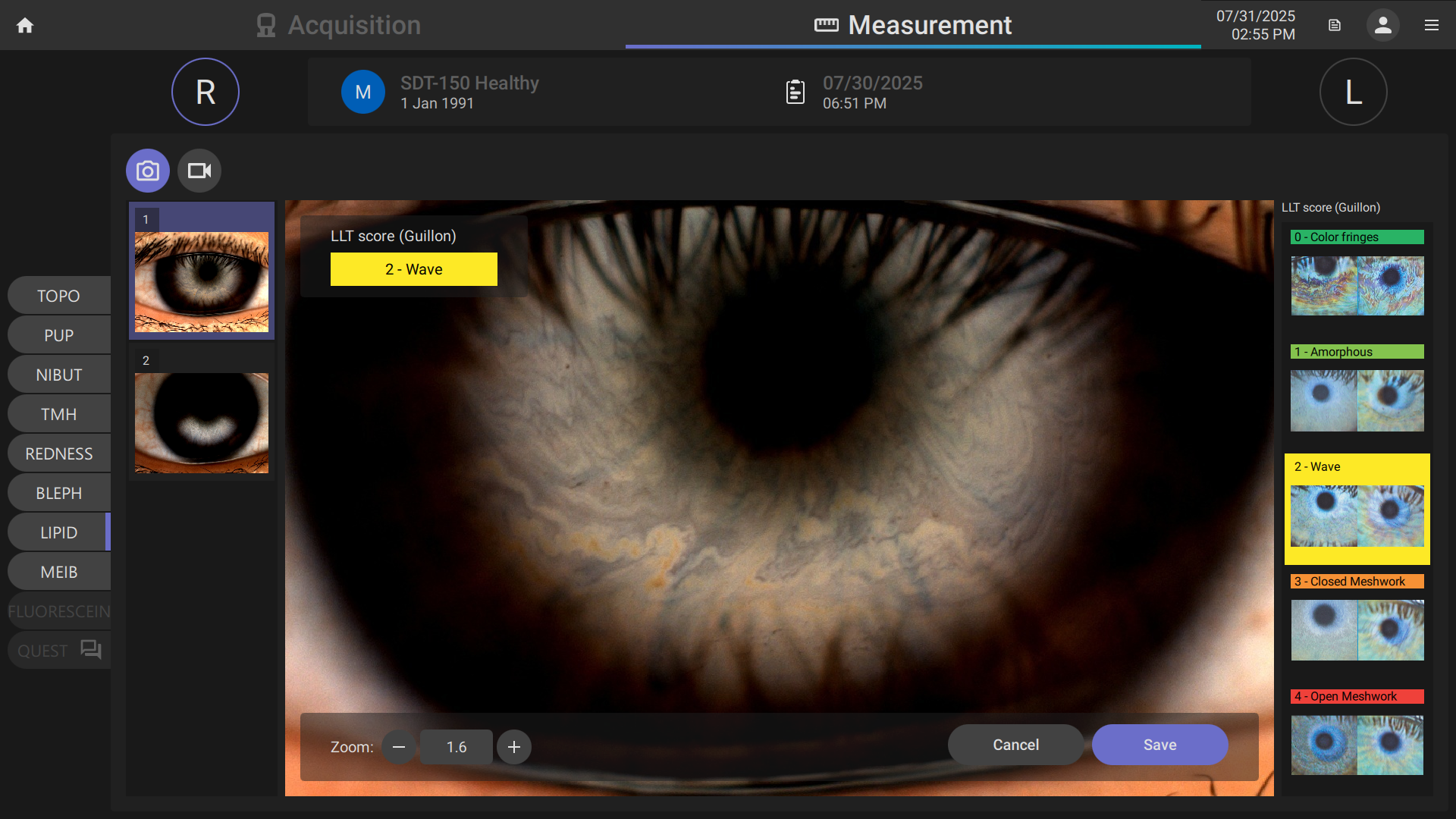
1. Guillon, M., 1998. Use of the Tearscope Plus in the routine contact lens fitting practice. Contact Lens and Anterior Eye, 21(Suppl 1), pp.S31-S40.
2. Pult, H. and Riede-Pult, B.H., 2012. Non-contact meibography: Keep it simple but effective. Contact Lens and Anterior Eye, 35(2), pp.77-80.
3. Efron, N., 1998. Grading scales for contact lens complications. Ophthalmic and Physiological Optics, 18(2), pp.182-186.
4. Jenvis, D.R., et al., 2007. The development and validation of the Jenvis Dry Eye Questionnaire. Optician, 233(6091), pp.16-21.
Note: The information contained on this website is intended for healthcare professionals. Not all products, services, or offers are approved or offered in every market, and products vary from one country to another. Contact your local distributor for country-specific information and availability.


