SP-1P

Easy-to-use automated endothelial analysis

The SP-1P photographs and displays analysed data by simply touching the image of the pupil on the control panel.
All functions, including alignment, photography and analysis are performed automatically.
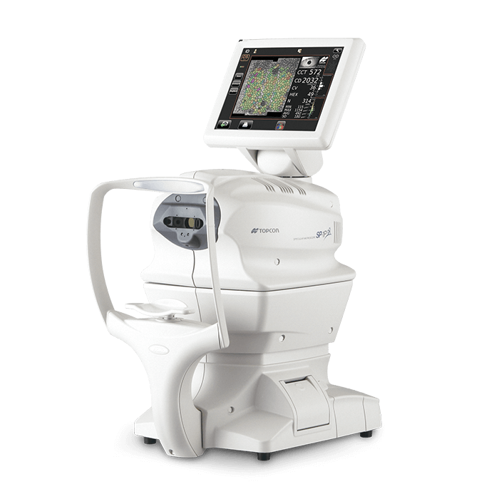
SP-1P

Key Features

  • Robotic Specular Microscope
  • Rapid and simple control by finger touch
  • Full 360° tlit of monitor
  • Flexible and Space saving layout
  • Panorama Photography Function
17 Fixation Points and Panoramic Function
17 fixation points distributed across the central, mid-peripheral and peripheral cornea offer multiple capture possibilities. In addition, the panoramic function covers a large, central corneal area for more complete evaluation of the patient’s endothelial condition*.

* The analysis of combined images is available after photographing a minimum of 2 areas (centre and either adjacent nasal or temporal).
Comprehensive Analysis Software
Key corneal metrics such as corneal thickness (CCT), cell density (CD), coefficient of variation (CV) and percentage of cell hexagonality (HEX), are clearly displayed on the results screen. Simply touch the screen to select the polymorphic or pleomorphic distribution histograms.
Analysis Report
The report can be exported as an image file.

Note:  The information contained on this website is intended for healthcare professionals. Not all products, services, or offers are approved or offered in every market, and products vary from one country to another. Contact your local distributor for country-specific information and availability.
Downloads

Request more information today

Whether you have product questions or are ready to purchase, we are standing by to help.
Fill out the form below and someone from our team will be in touch soon.