
A NEW ERA
BEGINS HERE
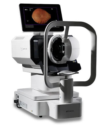
ULTRA-WIDEFIELD FUNDUS IMAGING. AUTOMATED, TRUE COLOUR, PRECISION. EFFORTLESS EXPERIENCE.
GAIA
Retinal Slit Scanner
SIMPLE OPERATION
FULLY AUTOMATED IMAGING, EFFORTLESS CAPTURE.
True Colour (RGB)
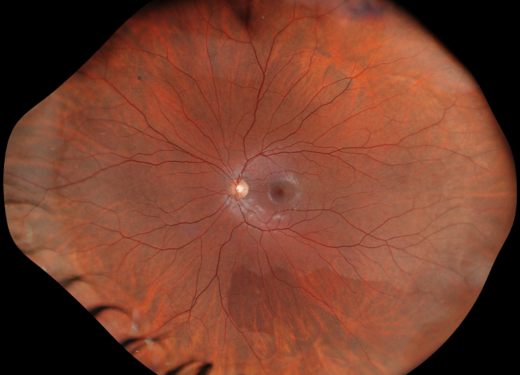
Utilising red, green and blue LED light sources (RGB), GAIA captures ultra-widefield retinal images in true colour *
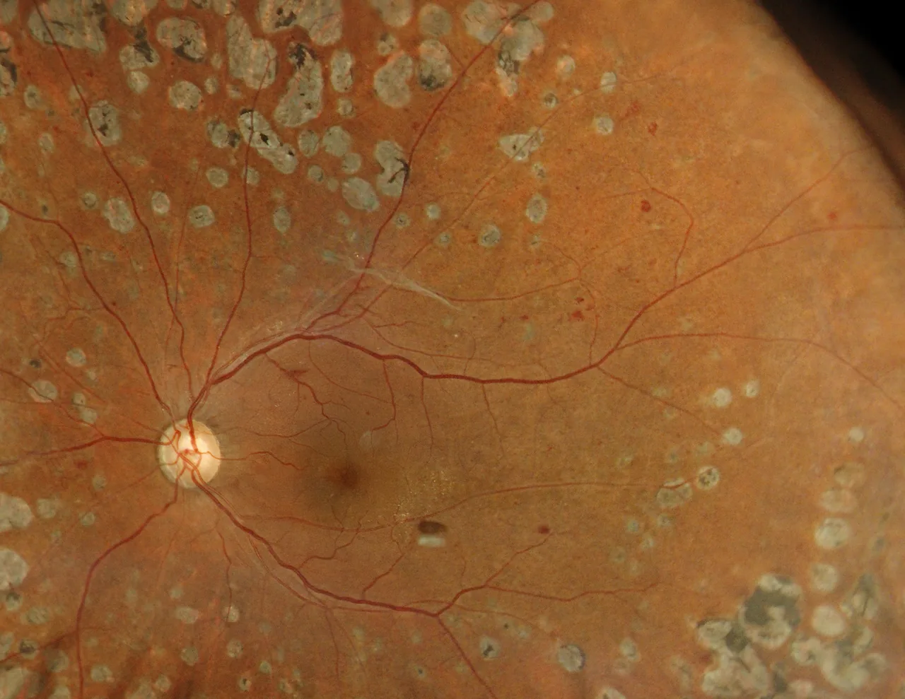
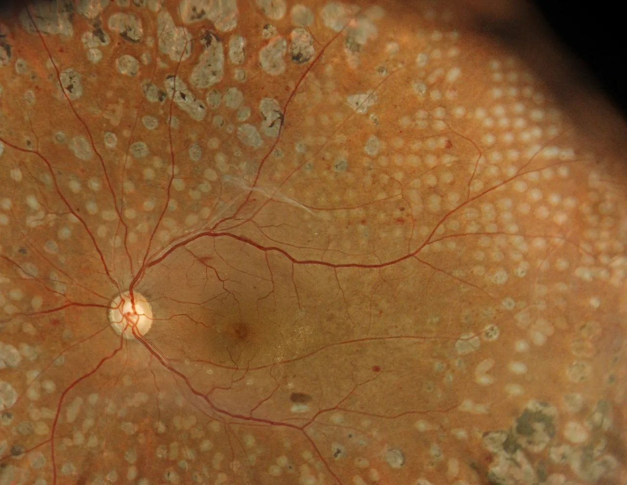
Diabetic Retinopathy — Courtesy: Dr. Shunsuke Osawa, MIE Eye Clinic
* True, full colour fundus image simultaneously captured with white light, 24-bit colour.
Slit-Scan Technology
Our cutting-edge slit-scanning technology utilizes a structured slit-light illumination combined with a high-speed rolling shutter mechanism. This method effectively blocks scattered light, allowing the system to achieve true colour ultra-widefield fundus images even through pupils as small as φ2.0mm without pharmaceutical dilation.
Key Features
-
 Capture more with 209°*ultra-widefield of view
 -
Improved patient comfort
-
Slit-scanning technology enables the acquisition
of high-quality ultra-widefield images -
Seamless integration
for efficient interoperability -
Clinical confidence with true colour imaging
-
Space-saving
-
Effortless operation, auto-alignment and capture
*209° is an estimated value derived from a visual angle of 150° (GAIA), based on the visual-angle-to-eye-angle relationship described in the following article.
Xincheng Yao, Devrim Toslak, Taeyoon Son, Jiechao Ma. Understanding the relationship between visual-angle and eye-angle for reliable determination of the field-of-view in ultra-wide-field fundus photography.
Biomedical Optics Express, 2021 Sep 30;12(10):6651-6659
Connectivity
With flexible export options via shared network folders or standard DICOM, GAIA adapts easily to a wide range of clinical environments. When connected to IMAGEnet7, it integrates seamlessly into existing workflows and creates a unified patient record across imaging modalities.
 USB Flash Drive
 GAIA
GAIA
 IMAGEnet7
 DICOM server
(Image storage)
Specifications
- Types of image-capturing
- Colour, Infrared (IR)*
- Resolving power on fundus
- Colour image-capturing: 20 lp/mm or more
- Measuring range for the dioptric power
- -20 D to +20 D
- Photographable diameter of pupil
- φ2.0mm or more (Confirmed with model eyes)
- Angular field of view
- 150° (Diameter of pupil φ2.5mm or more), 120° (Diameter of pupil φ2.0mm)
- Auxiliary functions
- Auto-alignment, Auto-focus, Auto-shoot
- Weight
- 31kg
- Dimensions
- 334 - 646 (W) × 549 - 573 (D) × 672 - 702mm (H)


44th Congress of the ESCRS 2026
GAIA will be showcased for the first time internationally at ESCRS 2026.
- Event
- ESCRS 2026
- Date
- 11-15 September, 2026
- Venue
- Excel London Royal Victoria Dock,1 Western Gateway, London E16 1XL
Discover GAIA at booth A. 150

Note: The information contained on this website is intended for healthcare professionals. Not all products, services, or offers are approved or offered in every market, and products vary from one country to another. Contact your local distributor for country-specific information and availability.

