
A NEW ERA
BEGINS HERE
ULTRA-WIDEFIELD FUNDUS IMAGING. AUTOMATED, TRUE COLOUR, PRECISION. EFFORTLESS EXPERIENCE.
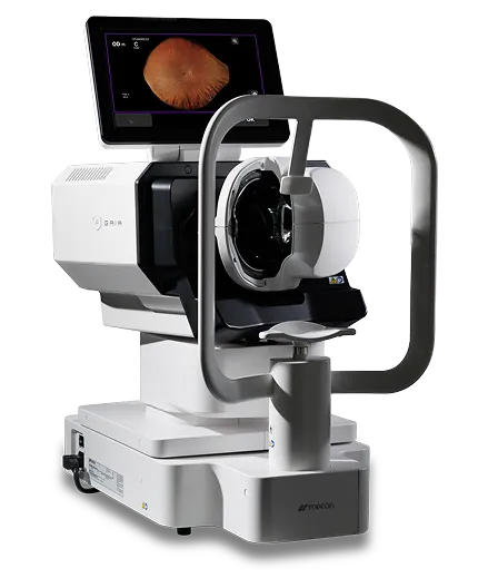
GAIA
Retinal Slit Scanner

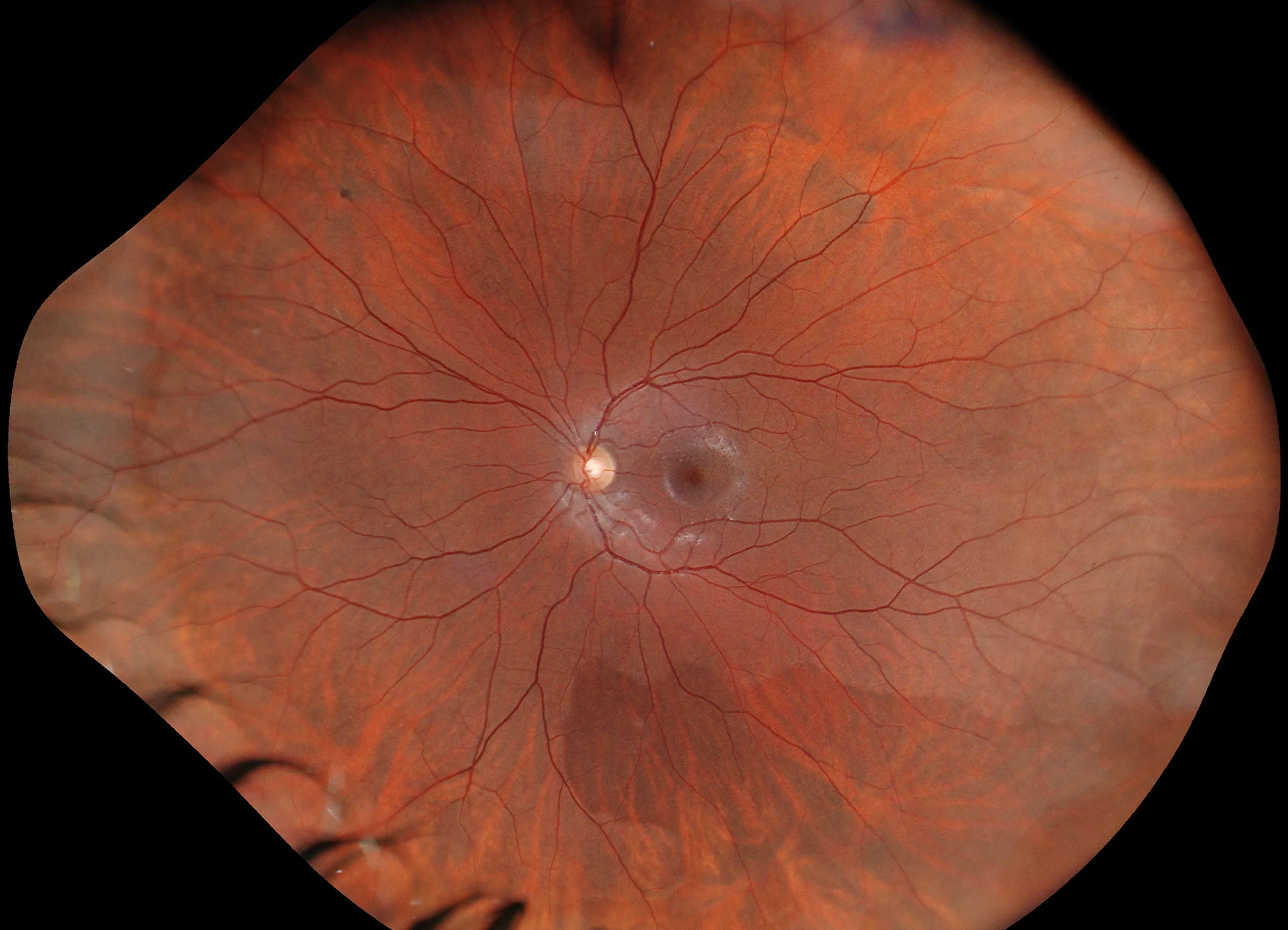
209°*
CAPTURE MORE, EFFORTLESSLY.
*209° is an estimated value derived from a visual angle of 150° (GAIA), based on the visual-angle-to-eye-angle relationship described in the following article.
Xincheng Yao, Devrim Toslak, Taeyoon Son, Jiechao Ma. Understanding the relationship between visual-angle and eye-angle for reliable determination of the field-of-view in ultra-wide-field fundus photography.
Biomedical Optics Express, 2021 Sep 30;12(10):6651-6659
SIMPLE OPERATION
FULLY AUTOMATED IMAGING, EFFORTLESS CAPTURE.
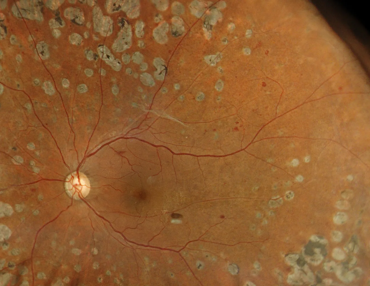
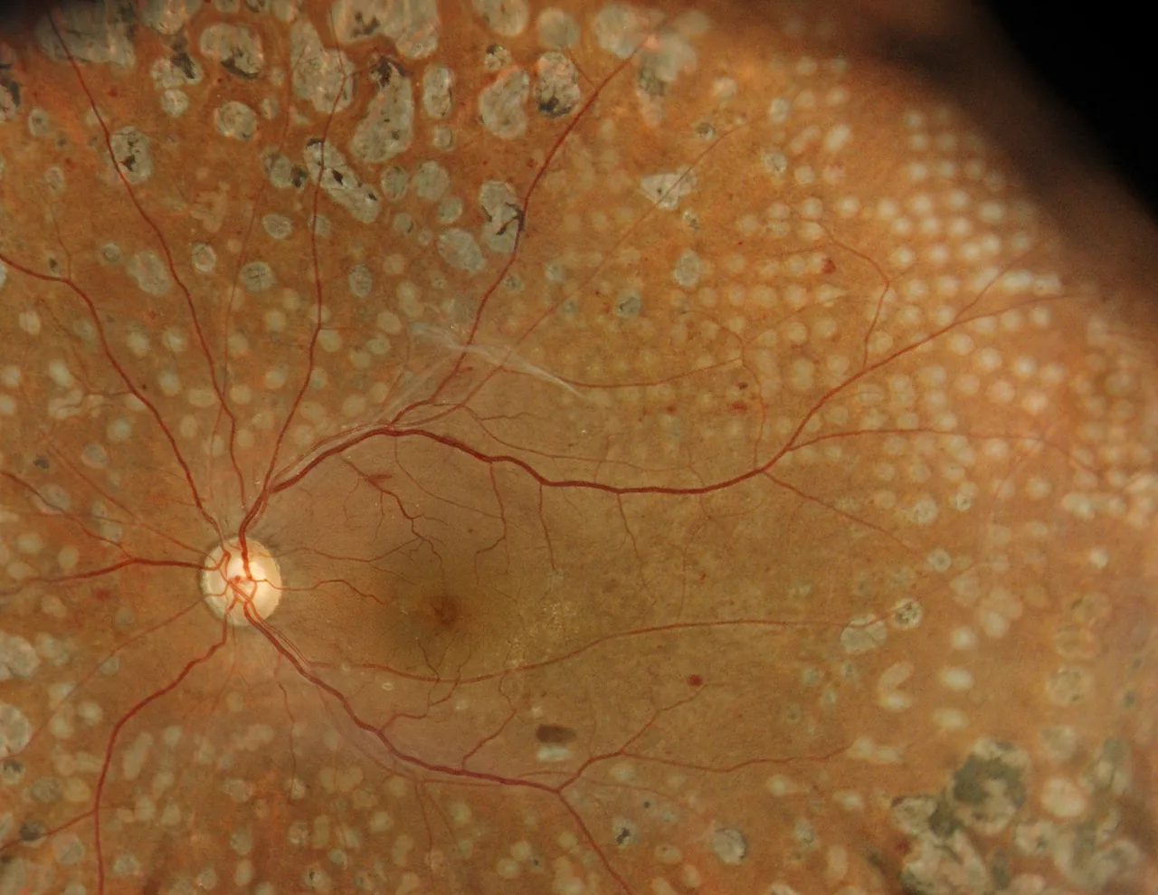
True Colour (RGB)
Utilising red, green and blue LED light sources (RGB),
GAIA captures ultra-widefield retinal images in true colour *
Diabetic Retinopathy — Courtesy: Dr. Shunsuke Osawa, MIE Eye Clinic
* True, full colour fundus image simultaneously captured with white light, 24-bit colour.
Slit-Scan Technology
Our cutting-edge slit-scanning technology utilizes a structured slit-light illumination combined with a high-speed rolling shutter mechanism. This method effectively blocks scattered light, allowing the system to achieve true colour ultra-widefield fundus images even through pupils as small as φ2.0mm without pharmaceutical dilation.
Key Features
-
 Capture more with 209°*ultra-widefield of view
 -
Improved patient comfort
-
Slit-scanning technology enables the acquisition
of high-quality ultra-widefield images -
Seamless integration
for efficient interoperability -
Clinical confidence with true colour imaging
-
Space-saving
-
Effortless operation, auto-alignment and capture
*209° is an estimated value derived from a visual angle of 150° (GAIA), based on the visual-angle-to-eye-angle relationship described in the following article.
Xincheng Yao, Devrim Toslak, Taeyoon Son, Jiechao Ma. Understanding the relationship between visual-angle and eye-angle for reliable determination of the field-of-view in ultra-wide-field fundus photography.
Biomedical Optics Express, 2021 Sep 30;12(10):6651-6659
Connectivity
With flexible export options via shared network folders or standard DICOM, GAIA adapts easily to a wide range of clinical environments. When connected to IMAGEnet7, it integrates seamlessly into existing workflows and creates a unified patient record across imaging modalities.
 USB Flash Drive
 GAIA
GAIA
 IMAGEnet7
 DICOM server
(Image storage)
Specifications
- Types of image-capturing
- Colour, Infrared (IR)*
- Resolving power on fundus
- Colour image-capturing: 20 lp/mm or more
- Measuring range for the dioptric power
- -20 D to +20 D
- Photographable diameter of pupil
- φ2.0mm or more (Confirmed with model eyes)
- Angular field of view
- 150° (Diameter of pupil φ2.5mm or more), 120° (Diameter of pupil φ2.0mm)
- Auxiliary functions
- Auto-alignment, Auto-focus, Auto-shoot
- Weight
- 31kg
- Dimensions
- 334 - 646 (W) × 549 - 573 (D) × 672 - 702mm (H)


